Summer Undergraduate International Research Experience

.png)
Week 7
-
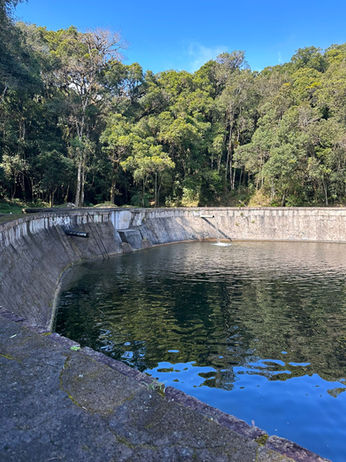
A student of Alexandre who works from Sao Paulo came to Curitiba this week to do some field work. Most of the lab studies crustaceans, and one in particular: Aegla schmitti. These crustaceans are mainly active in the winter, when waters are coldest. This student, Isabela, came along to collect aeglas for her Master's project. I was able to tag along for the day with them, and we went to Piraquara, a city where the natural waters come from. We explored this huge natural area where pools and streams of water gathered all types of animals. They had laid traps the day before, pieces of liver in plastic bottles with one-way entrance. I got to see and hold live aegla for the first time, which was exciting. They are funny-looking creatures that mostly walk backwards. I also got to see a lot of aquatic insects and other animals: mayfly larvae of all types, dobsonflies, caddisflies, stoneflies and a ton of tadpoles. I sifted a ton of leaf litter in a stream and was able to get about a dozen of aeglas (and the occasional accidental insect). It was so fun to participate in the field work and to help with Isabela's project.
For the rest of the week, I focused on my CT scans, and getting familiar with the program I would be using to analyze those. It was incredible to see a bunch of black and white horizontal slices become a full femur model.
Sifting crustaceans
Sifting crustaceans The week started off with field work at the EvoMorfo lab. Even though I had a lot to get done in my research, I knew there would not be another opportunity to explore the natural water sources of Curitiba in a city called Piraquara. On the coldest times of the year they collect the animal most of the lab studies: Aegla schmitti (see Fig. 1). Needless to say the weather was very cold, and the water even more. Wearing my humble sneakers and three layers of clothing, I was ready to help them find some aeglas. After checking on the liver traps set up the day before, we decided it would be best to sift on the spring waters. Knee-deep into the coldest water I ever stepped in, I sifted through leaf litter at the bottom of the waters. For some reason I seemed to have a talent for finding them because we ended up with a dozen of small aeglas and tons of aquatic insects. Doing field work remind me of why I love being a biologist. Something about finding your place in nature, of being an observer and getting to marvel at the intricate relationships of living things in a given environment. That was honestly the highlight of my week. The rest of the time I spent in the lab analyzing CT scans, looking through thousands of images and segmenting them into different parts to be quantified. A big thing that happened this week was figuring out there was no possible way to segment the apodeme (which is the part that links cuticle to muscle). My original idea was to use apodeme as a proxy for strength, being that the bigger connection from cuticle to muscle the stronger the force exerted when closing or opening the leg. Since most of our specimens are pretty old, this would be a safe bet since apodemes tend to be very resistant, much like the cuticle and unlike the muscles. Since Alexandre mostly worked with CT scans of crustaceans, which have easily visible and segmentable apodemes, we thought the same would be true for leaf-footed bugs. Turns out that the apodeme is so entrenched in the muscles that it is very hard to see them using micro-CT. Maybe with a better resolution (such as a synchotron) or staining, they would be more visible, but there is no way to know. We figured I can keep segmenting and see whether we find a big difference in the muscle density of different species (indicating deterioration) and if we do, we will go from there.


